Independent medical education supported by Novartis
Videos
Interview:
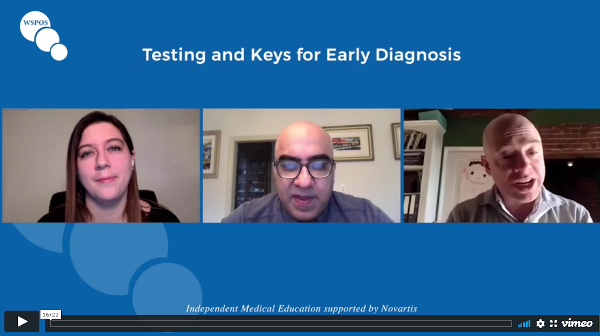
Testing and Keys for Early Diagnosis
Interview:
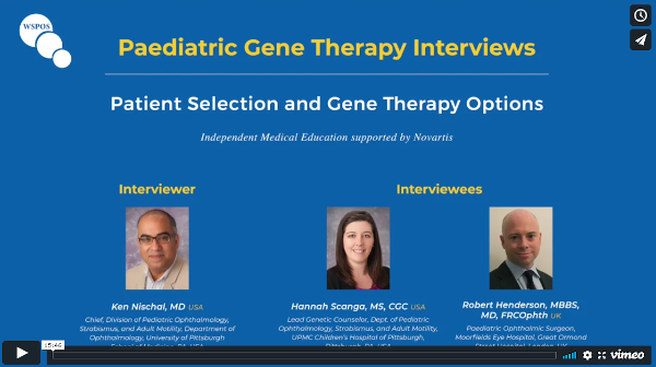
Patient Selection and
Gene Therapy Options
Interview:
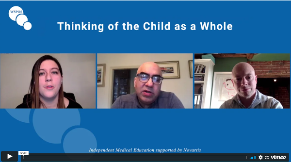
Thinking of the Child as a Whole
Podcasts
Podcasts
Episode 1:
Testing and Keys
for Early Diagnosis
with Ken Nischal, Robert Henderson and Hannah Scanga
In the first episode of the WSPOS paediatric gene therapy podcast Drs. Ken Nischal, Robert Henderson and Hannah Scanga talk about genetic testing and keys for early diagnosis of inherited retinal diseases. To hear more on paediatric gene therapy, listen to the other episodes in this series. Independent medical education support provided by Novartis.
with Ken Nischal, Robert Henderson and Hannah Scanga
In the second episode of the WSPOS paediatric gene therapy podcast Drs. Ken Nischal, Robert Henderson and Hannah Scanga discuss patient selection and gene therapy options. To hear more on paediatric gene therapy, listen to the other episodes in this series. Independent medical education support provided by Novartis.
with Ken Nischal, Robert Henderson and Hannah Scanga
In the last episode of the WSPOS paediatric gene therapy podcast Drs. Ken Nischal, Robert Henderson and Hannah Scanga talk about the importance of treating the child as a whole. To hear more on paediatric gene therapy, listen to the other episodes in this series. Independent medical education support provided by Novartis.